


A history of the microscope in ten objects
Early days
Object One: Compound microscope
There is no precise date for the invention of the microscope.
Optical lenses, including magnifying glasses and spectacles, have been used since ancient times to help enlarge small details. But it was not until the invention of the compound microscope in the 1600s that people were able to see the world that is invisible to the naked eye.
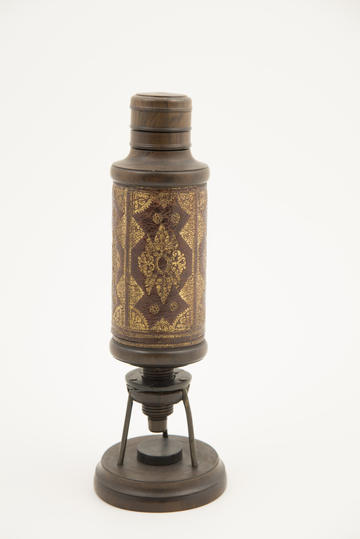
Our first object is a compound microscope dating from c.1670-80. It is a rare survival from this period of early discovery and use. Because compound microscopes use two or more lenses – rather than just one – to enlarge specimens, they achieve higher rates of magnification than a magnifying glass alone.
The user of this microscope would have placed their specimen on the small tripod base and observed it through the eyepiece at the top. Much of what they could see depended on lighting conditions: some used sunlight to illuminate their specimens, while others directed the light of an oil lamp through water to produce brighter, more concentrated beams.
Alongside their practical uses, the decorative value of early microscopes was important. Notice how the tube of this compound microscope has been decorated with a gold patterned leather, emulating the binding of an expensive book.
It would have been a prized possession for those who could afford such an instrument.
Members of the early Royal Society (the oldest national scientific society in the world, founded in 1660 to promote research into natural knowledge) were interested in exploring the possibilities of the microscope.
Robert Hooke, the Society’s laboratory assistant, was tasked with conducting a range of experiments with the microscope to test its capacity and function. Christopher Cock, the instrument maker who created this compound microscope, also made instruments for Hooke.
What Hooke saw when he looked down his compound microscope will be revealed in our second object: Robert Hooke’s Micrographia.
The bestseller
Object Two: Micrographia, Robert Hooke, 1665
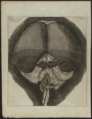
Hooke's Micrographia: The face and eyes of a drone fly
Robert Hooke (1635-1703) was a natural philosopher who was appointed as the Royal Society’s first Curator of Experiments in 1662, two years after the Society was founded.
He demonstrated experiments to the Society’s first Fellows, including ones using the microscope.
Hooke published the results of these experiments in his book Micrographia, the first official Royal Society publication, printed in 1665.
Micrographia was an instant bestseller.
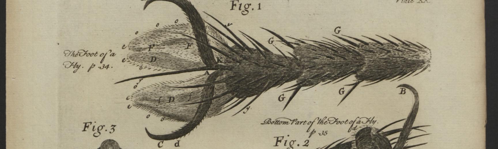
Readers were particularly captivated by the 38 copperplate engravings depicting everyday objects, including insects and plant materials, in breath-taking detail.
Most famous of all is Hooke’s flea, which transformed a tiny household pest into a huge, armoured creature.
Hooke’s bitter feud with Sir Isaac Newton (1643-1727) over alleged plagiarism, together with his own grouchy temperament, caused Hooke’s reputation to fade quickly in the 1700s.
However, the publication of an abridged edition of Micrographia in 1745 renewed his popularity amongst a new generation of microscopists. The edition focused mostly on reprinting his engravings, images which continue to delight readers to this day.
A microscope in the hand
Object Three: Leeuwenhoek replica microscope
Across the channel, the Dutch cloth merchant Antonie van Leeuwenhoek (1632-1723) was making his own microscopes. These small, single-lens instruments look dramatically different from the microscopes used by Robert Hooke (see object one).
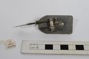
Object three is a 1933 replica of a style of microscope Leeuwenhoek first designed in the 1670s. To magnify specimens, it uses one small lens no bigger than a lentil. In fact, lens comes from the Latin word lens which means 'lentil'.
To use this microscope, Leeuwenhoek attached specimens to the pin and held the instrument up to his eye. This is a very different experience from using a compound microscope, where the viewer is physically separated from the specimen by the length of the microscope’s body.
How would you prefer to look at a spider? By peering down a tube like Robert Hooke, or holding it up to your eye like Leeuwenhoek?
Leeuwenhoek ground the lenses himself to make small, hand-held devices which achieved impressive levels of magnification, allowing him to observe smaller lifeforms than had ever been seen before.
He was one of the first people to observe what we know today to be red blood cells, sperm, and bacteria. He called these organisms 'dierken', which means animalcule or 'little animal' in Dutch.
From 1673, Leeuwenhoek wrote regularly to the Royal Society to describe what he had seen.
Robert Hooke often tried to reproduce Leeuwenhoek’s experiments. But this was not always easy, as Leeuwenhoek was deeply secretive about his method for making lenses. Leeuwenhoek also made a new microscope for each of his experiments. This made his observations difficult for others to replicate exactly.
Despite these challenges, Leeuwenhoek enjoyed a reputation as the leading microscopist during the 1700s, eclipsing Hooke in his authority and influence. He donated his microscopes to the Royal Society after his death.
Microscopes in your pocket
Object Four: Screw-barrel Microscope
Early microscopes were difficult to use and the images they produced could be distorted and confusing.
Users struggled to find the right lighting conditions for illuminating their specimens, and lenses were affected by aberrations which blurred the detail and colour of images (see object eight).
By the end of the 1600s, these frustrations had undermined the instrument's popularity in some circles.
In an address to the Royal Society in 1692, Robert Hooke identified Leeuwenhoek as the 'single votary' (advocate) of the microscope, stating,
besides whom, I hear of none that makes any other Use of that Instrument, but for Diversion and Pastime, and that by reason it is become a portable Instrument, and easy to be carried in one's Pocket.1

Our fourth object - the screw-barrel microscope - is an example of these new 'portable instruments’.
The screw-barrel microscope is an early design for a pocket microscope, an immensely popular model in England during the 1700s and 1800s. It could be taken apart and stored neatly in its case, meaning people could carry it with them and use it anywhere.
Once assembled, it functioned as a handheld microscope that the user focused by adjusting the barrel and held up to their eye, making it similar to Leeuwenhoek's design (see object three).
The light, portable design is very different from the bulky compound microscope which would have been used indoors (see object one).
The success of portable instruments like the screw-barrel microscope reflects the growing popularity in the 1700s of studying the natural world, especially botany (the study of plant life), entomology (the study of insects), and marine science (the study of oceanic life).
Women made important contributions to these studies, participating within the broader networks in which enthusiasts traded observations and specimens – often by post (see object ten).
Hooke’s criticism of the broadening use of the microscope represents an early questioning of what science should be and who should participate in it.
His words have inspired historians of science to view the 1700s as a time when the microscope was used for popular entertainment, rather than purely scientific enquiry. However, the fashion for field microscopes encouraged more people to examine the natural world, creating a culture of curiosity in which observations were conducted, shared, and reproduced.
Within these activities, the line between scientific enquiry and ‘diversion and pastime’ was by no means clear, with each supporting the other.
Nonetheless, instructing new audiences on how to use more specialised models remained a barrier to the instrument’s popularity.
If the microscope was to become an accessible instrument, a simple guidebook was needed (see object five).
Anything you can do
Object Five: The Microscope Made Easy by Henry Baker
Henry Baker (1698 – 1774) was a natural philosopher and antiquarian who forged a career in popularising the microscope.

The Microscope Made Easy … the fifth edition (Henry Baker, 1769)
In 1742, he published The Microscope Made Easy, which contained detailed instructions on how to use different microscopes, and examples of experiments to conduct. This included extended reprints of Leeuwenhoek's observations, passages from Hooke's Micrographia, and descriptions of Baker's own experiments with the microscope.
Baker believed that discoveries made by the microscope had been limited by the practical difficulties of using them. Microscopes were expensive, and even those who could afford one often struggled to see anything clearly.
In the preface to The Microscope Made Easy, Baker wrote how
‘many have been frightened from the Use of [the microscope], by imagining it required great Skill in Optics, and Abundance of other learning, to comprehend it to any Purpose'.
However, using the microscope could be easy: all one needed were
'good Glasses [microscopes], good Eyes, a little Practice, and a common Understanding'
And a copy of Baker’s book, of course.2
Baker's work supercharged the popularity of the microscope in the 1700s.
The Microscope Made Easy was hugely successful, running through five editions and undergoing translation into several languages, including Dutch and French.
It provided diagrams which showed the different types of microscopes available alongside step-by-step instructions on how best to use them.
Baker rarely cited the observations of female microscopists in his work, though he did rely heavily upon the private collections of women when searching for particular specimens to observe.
Baker’s book was also designed to help people select the right instrument for their personal interests. An expanding range of specialised instruments were available in the 1700s, including field microscopes (see object four) and solar microscopes (object six).
As a result, The Microscope Made Easy profited from, and encouraged, the growing culture for popular science in England during the 1700s.
Putting on a show
Object Six: Solar Microscope
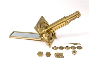
The invention of the solar microscope in the mid-1700s transformed the experience of using the microscope.
Much like the camera obscura, solar microscopes used sunlight to project specimens onto a wall.
Suddenly, people no longer saw fleas wriggling at the end of their microscope: their image crawled across the wall a hundred times larger than life.
The effect was akin to an early form of cinema, shocking and amusing its audience.
In The Microscope Made Easy (see object five), Henry Baker wrote how
This [solar] Microscope is the most entertaining of any; and perhaps the most capable of making Discoveries.
It allowed people to view specimens together and discuss what they saw, and by
discoursing on what lies before them, may be able better to understand one another, and more likely to find out the Truth, than when, in other Microscopes, they must peep after another.3
This group experience complemented an emerging culture of popular and sociable science in Georgian England.

Illustration of the solar microscope in use, from Martin Ledermuller’s Amusemens Microscopiques (Microscopic Amusements), 1768
Solar microscope relied on the light of the sun, meaning a cloudy day or the movement of the sun across the sky all frustrated its ability to put on a show, rain or shine.
Nonetheless, solar microscopes transformed microscopy from a private pastime into collective entertainment.
Its showmanship and spectacle helped observers discuss experiments and verify their findings together, providing education through entertainment.
Style over substance?
Object Seven: George III's Microscope

This microscope is fit for a king.
Sumptuous, ornate, and decked in silver, it was made for King George III in 1761 by George Adams, the official instrument maker for the king.
Don’t be too distracted by its decorative extravagance: nestled between cherubs and floral details are two functional microscopes, one simple (which uses a single lens) and one compound (which magnifies using two or more lenses).
Can you spot them? (One black eyepiece is above the heads of the figures at the top, and the other is to the left of the figures’ feet).
This microscope was designed to communicate more than just kingly wealth. It proclaimed George III's status as an enlightened monarch who supported scientific investigation.
Nonetheless, historians have hailed this instrument as 'history's most ludicrous microscope', arguing its design prioritised beauty over functionality.
The decorative accessories are certainly attractive, but their size and placement interfere with the user’s ability to conduct observations. As a result, this microscope looks more like an elaborate candlestick than it does a tool for scientific experimentation.
But when it comes to making microscopes, decoration and function have always been entwined.
Our first microscope is decorated with a gold patterned leather of no practical value (see object one).
Hooke's engravings in Micrographia were as much celebrated for their artistic beauty as they were for their scientific knowledge (see object two)
And the ivory of the screw-barrel microscope (object four) and brass of the solar microscope (object six) served both practical and decorative purposes.
As a result, it is important not to draw too harsh of a line between artistic licence and scientific function – even for a microscope as extravagant as this one.
Coming into focus
Object Eight: Microscope Lenses
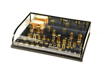
Before the 1800s, the images produced by microscopes were not always clear.
Early lenses could not focus the all colours (wavelengths) of light to a single point. They produced an optical effect known as chromatic aberration.
This meant that images were slightly blurred and distorted by a halo of colours, limiting the details early microscopes were able to reveal.
Our eighth object is a case of microscope lenses designed by Joseph Jackson Lister (1786-1869). Lister began studying optics in the mid-1820s, and worked with the optician William Tulley to design lenses that did not produce image aberrations. These new lenses (known as achromatic lenses) were hugely successful and became an industry standard for instrument makers.
Lister lenses changed the experience of looking through the microscope.
Like Henry Baker’s 1742 popular microscopy manual, The Microscope Made Easy (see object five), they helped make the microscope less confusing to use as images were clearer and colours more accurate.
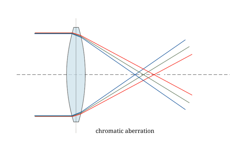
Chromatic aberration of a single lens causes different wavelengths of light to have differing focal lengths (CC BY-SA 3.0)
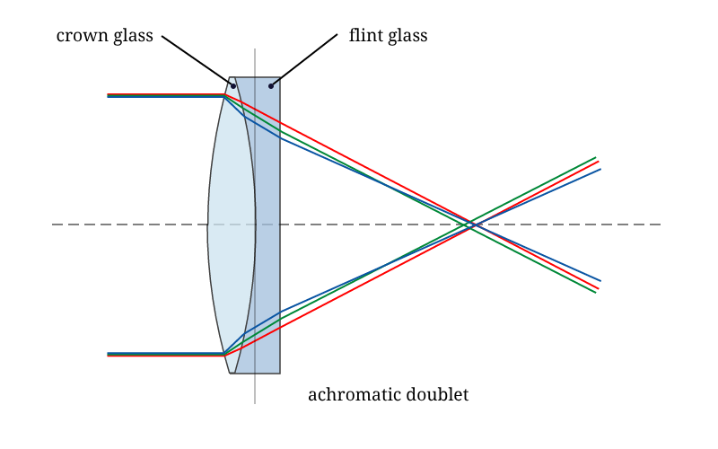
An achromatic doublet brings red and blue light to the same focus and is the earliest example of an achromatic lens (CC BY-SA 3.0)
While this marked an important moment in the development of the microscope, we should not assume that earlier microscopes were unable produce good images.
The work of Robert Hooke (object two), Leeuwenhoek (object three) and Henry Baker (object five) all demonstrate the capacity of microscopes in the 1600s and 1700s to produce meaningful discoveries.
However, the advancements made in lens technologies in the 1800s helped the microscope to produce more fine-tuned observations, supporting the growing popularity of microscopy as both science and entertainment in Victorian England.
The Listers were a family of firsts. Joseph Jackson Lister’s son, the surgeon Joseph Lister, is also celebrated in the history of medicine for developing the first antiseptic practices.
Microscopy on film
Object Nine: Photographic Microscope
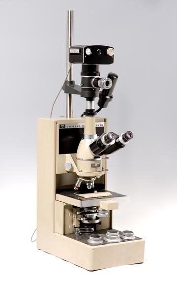
How can you capture the image you see when you look through a microscope?
Should you draw it, or should you describe it?
Can you convince others of the remarkable complexity of everyday objects?
How might you explain these sights to someone who has never used a microscope?
These questions have echoed throughout the microscope’s history.
In Micrographia, Robert Hooke responded by including powerful copperplate engravings which sought to recreate the view through the lens of his microscope (see object two).
He also included descriptions which compared the strange and unexpected sights he saw to images common to his readers. For example, the edge of a razor appeared
almost like a plow’d field, with many parallels, ridges, and furrows.4
Our ninth object is a photographic microscope from the 1980s which features a Kodak camera body.
The history of the microscope is closely related to the development of photography: advancements in optical knowledge produced new technologies for viewing and capturing images, and instrument makers were quick to incorporate these techniques into their models.
From the 1700s, microscopists used solar microscopes (see object six) and the camera lucida – an accessory which uses a prism to superimpose an image onto a drawing surface – to trace what they saw onto paper.
By the 1800s, microscopists began using photography to capture images directly – a technique known as photomicrography.
Photomicrography has allowed microscopists to communicate and disseminate their findings with increased precision.
This has had a particular impact on medicine: for example, Robert Koch used the technology in the late 1800s to photograph bacteria, supporting his discoveries in pathology.
Photomicrography can also be used to monitor a patient’s condition by documenting diseased cells and responses to treatment.
Today, scientists continue to use micrography to capture and share observations, demonstrating the importance of photographic microscopes as an agent of scientific communication.
It's in the mail
Object Ten: Collection of Microscope Stamps
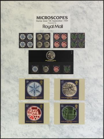
The final object in this history of the microscope is a collection of four stamps released in 1989 by the Royal Microscopic Society in celebration of their 150th anniversary.
The Society was founded in 1839 by a group of gentlemen including Joseph Jackson Lister (see object eight) and was granted a royal charter in 1866.
The History of Science Museum holds a significant collection of microscopes on loan from the Royal Microscopical Society, including several featured in this history: Leeuwenhoek’s replica microscope (object three), the solar microscope (object six), the Lister lenses (object eight), and the photographic microscope (object nine).
The images on the stamp depict key moments in the history of microscopy.
The 19p stamp depicts a snowflake magnified ten-times and the 27p stamp a blue-fly magnified five-times.
Both stamps evoke images from Robert Hooke’s Micrographia (see object two), which revealed the surprising complexity of tiny objects through its detailed illustrations – like those featured on the stamps.
The 32p stamp shows red blood cells magnified by 500 as they were seen by Lister and the founding members of the Society in 1839, celebrating the microscope’s contributions to medicine and microbiology.
And finally, the 35p stamp shows a microchip enlarged 600 times by an electron microscope, a model from the 1900s which uses electrons to magnify whole objects or surfaces, as opposed to slices.
The postal service has played an essential role in the development of microscopy. From the 1700s, growing networks of correspondence allowed enthusiasts to share observations, ideas, and advice.
Specimens were regularly requested and exchanged between correspondents, including botanical cuttings and small bottles of aquatic lifeforms. These circles of correspondence were particularly important to individuals who may have been excluded from formal cultures of collective science, including women and those who lived far away from urban centres.
Postal networks also connected the work of English microscopists to global currents of movement and trade.
Colonial merchants supplied male and female naturalists with specimens from across the world, which became jewels in their collections.
As a result, the postal service did not only support attempts to reproduce observations and advance natural knowledge. It also connected microscopy to projects of colonial expansion in which natural resources were extracted for private gain.
Made in an era of global connectivity and international trade, these Royal Mail stamps represent the importance of collaborative exchange in the development of microscopy.
Their function facilitates this conversation, while their design spotlights the images produced by the microscope, from Hooke’s early observations to the tiny technologies that power our own digital age.
Final thoughts
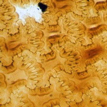
Exploring the history of the microscope in ten objects reveals the evolution of English microscopy from the 1600s to the present day.
From the screw-barrel microscope that could be carried in your pocket to the extravagant instrument that showed off George III’s wealth, the size and shape of the microscope can vary depending on its purpose.
The design of each microscope affected the experience of using it, contrasting the individual experience of using a Leeuwenhoek microscope against the collective theatre of the solar microscope.
And finally, the quality of lenses impacted the images produced by the microscope and shaped attempts to capture these views - from the magnificence of Hooke’s engravings in Micrographia to a special issue of Royal Mail stamps.
Throughout its history, the microscope has made important contributions to the development of scientific and medical knowledge.
Historians have traditionally focused on advances in germ theory and pathology during the 1800s, sidelining the activities of preceding centuries as simple forms of entertainment.
The microscopists of the 1600s and 1700s would not have seen their efforts in this way.
Men and women took great delight in using their instruments and were thrilled by the theatre of life it revealed.
But they did not consider this enjoyment to be incompatible with the advancement of knowledge.
Their approach to science is a reminder of how important curiosity and amusement are in making science accessible to all.
After all, the thrill of using the microscope to reveal a hidden world remains as enchanting today as it ever has been.
September 2026

